Abstract
3D Printing Optogenetic Interfaces
Optogenetics allows light to communicate with nerves. To enable this communication in the peripheral nervous system, nerve cuffs wrap around the nerve and hold a light source, usually a fiber optic cable or a micro light-emitting diode (µLED), to the nerve. To facilitate mouse research in different parts of the peripheral nervous system, a way to print nerve cuffs with 3D printers is being developed.
Why 3D print optogenetic interfaces?
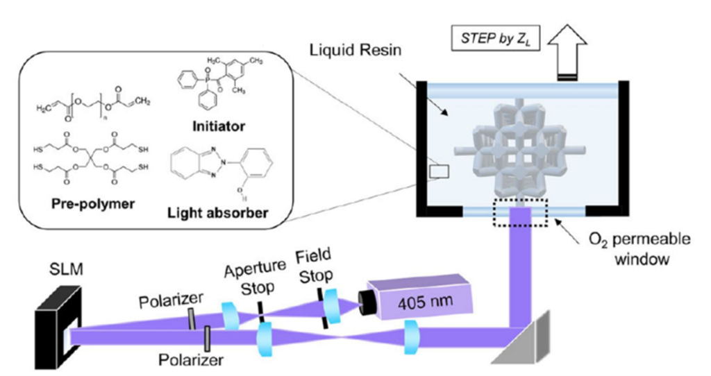
3D printers allow for the rapid iteration of different designs and give the ability to customize the design for specific anatomical geometries. Using a Digital Light Processing (DLP) 3D printer in the McLeod Lab at the University of Colorado Boulder, nerve cuffs can be 3D printed with biocompatible materials, using light to cure each layer. DLP 3D printers use a vat of liquid resin and a light to cure each layer of the part. As shown in Figure 1, the build plate steps up with each layer, allowing the light to polymerize a new layer. This allows for the design of intricate parts and fine details.
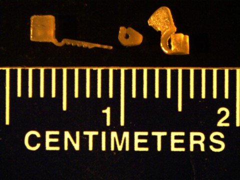
Below are some sample prints of optogenetic interfaces created using this 3D printer. These are potential designs for three different nerve cuffs.
What characteristics are important for nerve interfaces?
Material properties are important to consider when creating devices that will be implanted in the body. We are concerned about the following:
- Biocompatibility: Our materials should be non-cytotoxic and ideally, meet the guidelines given by ISO 10993
- Soft: Our material should have an elastic modulus ≤ 5MPa because this is similar to the elastic modulus of nerve tissue.4
- Tough: The material should be robust under forcep handling to allow for smooth implantation during surgery.
- High-Resolution Printing: The feature sizes must be smaller than 183µm as the Cervical Vagus Nerves in mice are approximately 183µm in diameter.5
What material should be used to print these interfaces?
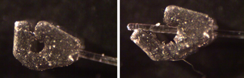
We are currently investigating using BioRes-Silicone from B9Creations. Figure 3 shows some nerve cuffs in this material created on the B9 Core 530J printer.
How do these interfaces work?
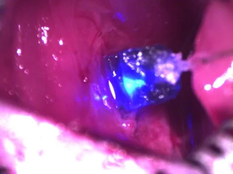
These interfaces are implanted around a nerve in a mouse, as shown in Figure 4. This study is focuses on the stimulation of the vagus nerve. These nerve cuffs were implanted around the left cervical vagus nerve in a mouse. Figure 5 shows the stimulation of the nerve using the light from a fiber optic cable inserted into a hole in the nerve cuff.
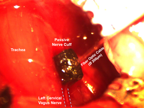
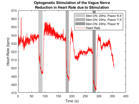
Stimulating the left cervical vagus nerve has been shown to reduce heart rate. As shown in Figure 6, the nerve cuff successfully stimulates the nerve when the light is on (gray rectangles), as indicated by the significant drop in heart rate.
What’s next in our research?
We will be redesigning the nerve cuff for µLED instead of a fiber optic cable. We will be implementing this improved system in various diseased mouse models where we believe vagus nerve stimulation to be therapeutic.
People
Weir Biomechatronics Development Laboratory
Current People
- Tyler R. Currie – PhD Student, CU Anschutz
- Arjun K. Fontaine, PhD – CU Anschutz
- Richard F. Weir, PhD – CU Anschutz
Past People
- Sam Littich, MS
Collaborations
McLeod Lab – University of Colorado | Boulder
- Robert R. McLeod, PhD
Posters & Conference Presentations
Currie, T.R., Mcleod, R.R., Weir, R.F.ff, Fontaine, A.K., (2024): Designing a 3D Printed Nerve Cuff for Optogenetic Vagus Nerve Stimulation in Mice. VA Research Day, 9/26/2024, Aurora, CO, September 2024. Accepted for Presentation.
Currie, T.R., Mcleod, R.R., Weir, R.F.ff, Fontaine, A.K., (2024): 3d Printing Optogenetic Interfaces. Neuro Engineering Mini Symposium, 4/29/2024, Aurora, CO, April 2024. Accepted for Presentation.
Currie, T.R., Mcleod, R.R., Weir, R.F.ff, Fontaine, A.K., (2023): 3d Printing Optogenetic Interfaces. Society for Neuroscience 2023, PSTR513, Optogenetics and Applications, 11/15/2023 8:00am, Washington DC, Nov. 2023. Accepted for Presentation.
Publications
Littich SF (2020) Design of a Reusable Elastomer Cuff for Stable in Vivo Imaging of Murine Vagus Nerve Axons. In: University of Colorado at Denver.
Funding
This work was funded in part through:
- NINDS R21 NS124313-01 (Weir, Fontaine): A 3D-Printed Nerve Cuff for 1-Photon Optogenetic Vagal Stimulation
- NIH NINDS R01NS118188(Weir, Caldwell, Gibson): Optimization of a Minimally Invasive Bidirectional Optogenetic Peripheral Nerve Interface with Single Axon Read-in & Read-out Specificity.
- Department of Veterans Affairs, Rehabilitation Research and Development Service, administered through the VA Eastern Colorado Health Care System – Rocky Mountain Regional VAMC, I21 RX003894-01 (Fontaine): Investigation of an Optogenetic Vagus Nerve Stimulation Device in an Animal Model of Post-traumatic Stress Disorder.
Patents
News & Media
References
- Uzcategui, A. C., Higgins, C. I., Hergert, J. E., Tomaschke, A. E., Crespo-Cuevas, V., Ferguson, V. L., Bryant, S. J., McLeod, R. R., & Killgore, J. P. (2021). Microscale Photopatterning of Through-thickness Modulus in a Monolithic and Functionally Graded 3D Printed Part. Small Sci, 1(3). https://doi.org/10.1002/smsc.202000017
- Littich SF (2020) Design of a Reusable Elastomer Cuff for Stable in Vivo Imaging of Murine Vagus Nerve Axons. In: University of Colorado at Denver.
- Uzcategui, A. C., Muralidharan, A., Ferguson, V. L., Bryant, S. J., & McLeod, R. R. (2018). Understanding and Improving Mechanical Properties in 3D printed Parts Using a Dual-Cure Acrylate-Based Resin for Stereolithography. Adv Eng Mater, 20(12). https://doi.org/10.1002/adem.201800876
- Guimarães, C. F., Gasperini, L., Marques, A. P., & Reis, R. L. (2020). The stiffness of living tissues and its implications for tissue engineering. Nature Reviews Materials, 5(5), 351-370. https://doi.org/10.1038/s41578-019-0169-1
- Stakenborg, N., Gomez-Pinilla, P. J., Verlinden, T. J. M., Wolthuis, A. M., D’Hoore, A., Farre, R., Herijgers, P., Matteoli, G., & Boeckxstaens, G. E. (2020). Comparison between the cervical and abdominal vagus nerves in mice, pigs, and humans. Neurogastroenterol Motil, 32(9), e13889. https://doi.org/10.1111/nmo.13889