Biomimetic Prosthesis-Orthosis Prototype
Testing a footplate to improve roll-over shapes in the gait-cycle
Biomimetic Prosthesis-Orthosis Prototype
Testing a footplate to improve roll-over shapes in the gait-cycle
Abstract
Biomechanical Testing of a Biomimetic Prosthesis-Orthosis Prototype
The human foot is made of uniquely shaped bones that form two arches. The main longitudinal arch begins at the hindfoot, ends at the distal articulations of the metatarsals (forefoot), and is tightened underneath by plantar fibers, tendons, muscles, and the plantar aponeurosis. The construction of the foot creates the windlass mechanism, a passive structure that stiffens the arc and assists in walking.1

(Retrieved from Ian Griffiths Sports Podiatry)
The forefoot provides stability and bears a significant amount of weight during push off in walking. Loss of the forefoot does not severely hinder performance of activities of daily living, but the mechanical changes of walking gait does impact the ease and safety of these tasks.2 Current bodies of research show that the function of the foot adapts to a loss of balance,3 abnormal tracking of the center of pressure (CoP),4 and asymmetries in gait biomechanics that can result in injury on the contralateral limb.5 In addition, disarticulation of the forefoot removes the support of the longitudinal arch, effectively neutralizing the effect of the windlass mechanism. In 2008, Ziegler-Graham et. al. estimated the prevalence of limb loss in the United States and reported that about 65% of amputations were of the lower limb (N > 1 million). Of the lower limb amputations, about 40% were of the phalanges.6 The team projects that these numbers will double by 2050. Additionally, those who sustain a partial foot amputation are likely to have a subsequent amputation that removes more than half of the foot, if not all of it.7
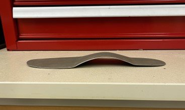
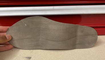
Based on the existing literature, it is still unclear how to characterize walking gait in individuals with partial foot amputations. Additionally, there is still a need for an effective intervention for partial foot amputations that can restore the mechanical advantage of the foot. This project aims to evaluate and optimize an existing prototype device from the Biomechatronics Development Laboratory at Anschutz Medical Campus. The device is a biomimetic prosthesis-orthosis hybrid that is made of a continuous piece of steel.
Figure 2: Top (a) and Side (b) views of the steel, biomimetic prosthesis/orthosis hybrid that was 3D printed in the Weir Biomechatronics Development Laboratory.

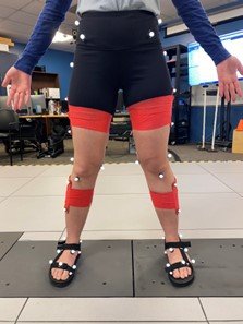
Motion capture data will be collected at the Interdisciplinary Movement Science Laboratory. We will use this data for rigid body inverse dynamics that inform us on how the device affects walking, the function of the foot, and design considerations.

People
Weir Biomechatronics Development Laboratory
Current:
- Project Lead: Jordan McClung
- Stephen Huddle, MS
- Stephanie Lorelli, MS
- Richard F. ff. Weir, PhD
Collaborations
University of Colorado, Anschutz Medical Campus
- Brecca Gaffney, Ph.D, Assistant Professor, Department of Mechanical Engineering
Posters & Conference Presentations
Publications
Funding
This work was funded in part through:
- Funds from the Department of Veterans Affairs, Rehabilitation Research and Development Service, administered through the VA Eastern Colorado Health Care System – Rocky Mountain Regional VAMC
Patents
News & Media
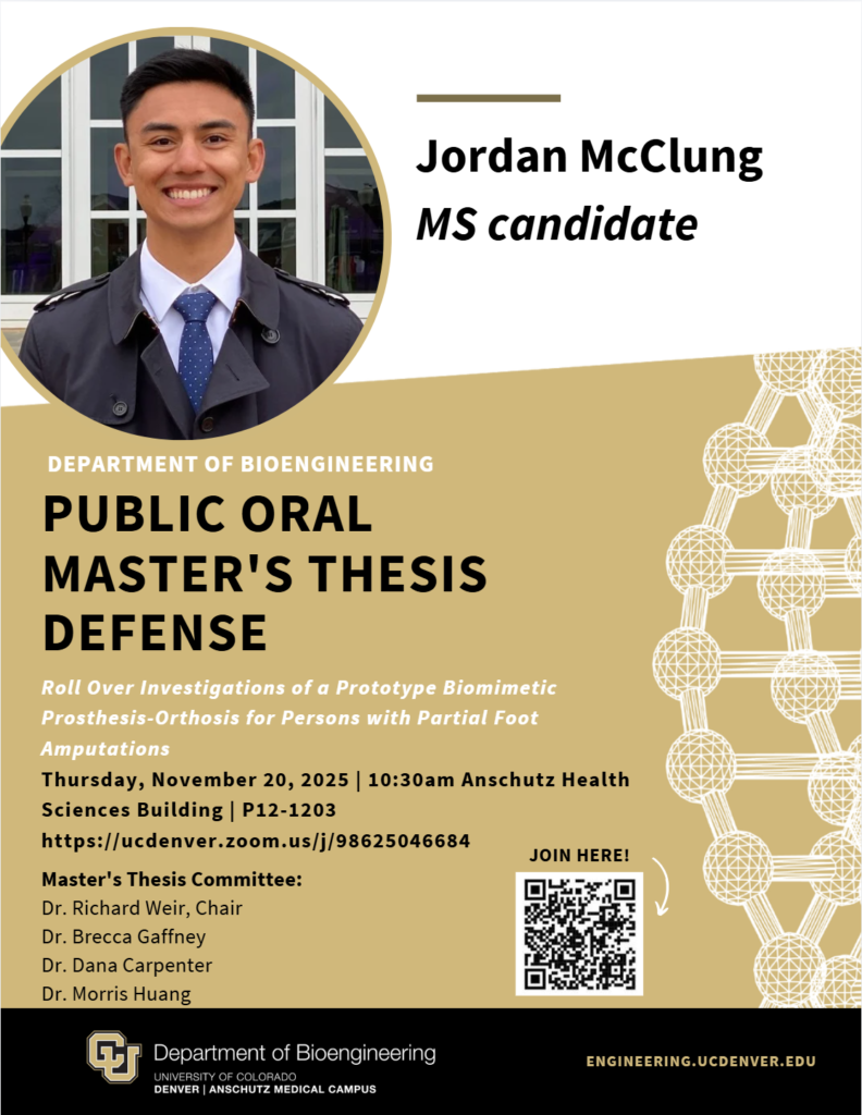
Jordan McClung defended his Master’s Thesis on November 20th, 2025!
References
- R. A. Mann and J. L. Hagy, “The Function of the Toes in Walking, Jogging and Running,” Clinical Orthop. Relat. Res., vol. 142, pp. 24–29, 1979.
- L.C. Feamster, “Exploratory Design and Evaluation of a Biomimetic Hybrid Prosthetic-Orthotic Intervention for Amputations of the Great Toe”. [Unpublished Master’s Thesis] University of Colorado, 2020.
- S. W. Chou, H.-Y. K. Cheng, J.-H. Chen, Y.-Y. Ju, Y.-C. Lin, and M.-K. A. Wong, “The Role of the Great Toe in Balance Performance,” J. Orthop. Res., vol. 27, pp. 549–554, 2009.
- R. A. Mann, N. K. Poppen, and M. O’Konski, “Amputation of the Great Toe: A Clinical and Biomechanical Study,” Clin. Orthop. Relat. Res., vol. 226, pp. 192–205, 1988.
- P. G. Adamczyk and A. D. Kuo, “Mechanisms of Gait Asymmetry Due to Push-Off Deficiency in Unilateral Amputees,” IEEE Trans. Neural Syst. Rehabil. Eng., vol. 23, no. 5, pp. 776–785, 2015.
- K. Ziegler-Graham, E. J. Mackenzie, P. L. Ephraim, T. G. Travison, and R. Brookmeyer, “Estimating the Prevalence of Limb Loss in the United States: 2005 to 2050,” Arch Phys Med Rehabil, vol. 89, pp. 422–429, 2008.
- D. P. Murdoch, D. G. Armstrong, J.B Dacus, T. J. Laughlin, C. B. Morgan, and L. A. Lavery, “The Natural History of Great Toe Amputations,” J Foot Ankle Surg, vol. 36, no.3 May, pp.204-208, 1997.